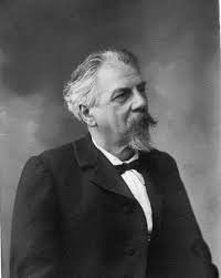
Snellen and his visual acuity chart

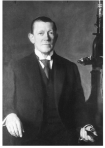
A day in an optometry clinic or vision camp doesn’t go without a visual acuity chart. Although there are various types of charts, the most common one is Snellen visual acuity chart. Herman Snellen (February 19, 1834 – January 18, 1908), a dutch ophthalmologist developed the chart in 1862 to measure visual acuity. Today, Jan 18 marks his death anniversary.
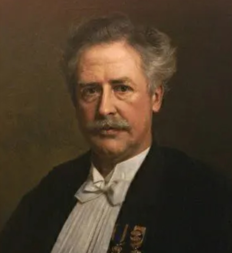
The chart has 11 lines of optotypes constructed as per strict geomteric rules. Each row of letters is assigned a ratio which indicates the visual acuity required to read it, and the ratio for the lowest line a person can read represents the individual’s visual acuity for that eye. A ratio less than 1 (for instance, 6/10) indicates worse-than-normal vision; a ratio greater than 1 (for instance, 6/5) indicates better than normal vision.

Want to learn how to make a visual acuity chart?

Conversely there are some controversies in using Snellen as a standard chart:

  1. The number of letters increases from top to bottom. This might cause visual crowding to some patients and reduce the visual acuity than the actual vision.
  2. The ratio difference between cosecutive lines is not similar and does not follow a particular order
  3. Spacing between the rows and letters is not similar or does not follow a linear or exponential order
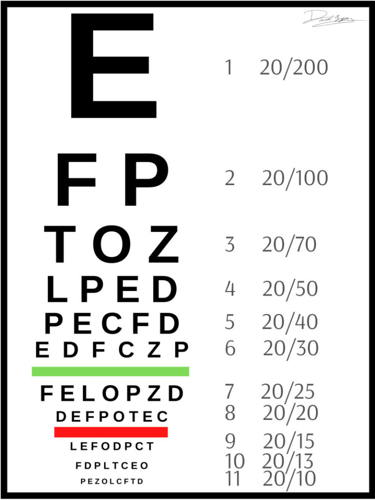
Another interesting invention of Snellen was designing a light hollow prosthesis along with Muller brothers in 1892. The shell prosthesis was originally placed over atrophic eye and used after enucleation. This caused serious discomfort for the patient. To avoid this problem, Snellen designed a hollow prosthesis that could fill the space and fix in the socket well. This has been a real success in those times.
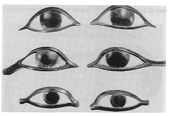
Herman Snellen (1834-1908) studied medicine in Utrecht and wrote a thesis under the guidance of Franciscus Donders (1818-1889). Yes, he was the Donders who introduced prismatic and cylindrical lenses for managing astigmatism and invented impression tonometer.

He invented numerous surgical procedures. These also include equipments related to entropion, ectropion, and trichiasis. Next time when you walk into an optical store and look at a big E on the top of a chart, remember the mastermind behind the chart is Snellen.
It’s Krause’s birthday!

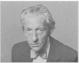
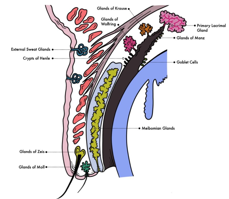
Karl Friedrich Theodor Krause (15 December 1797 – 8 June 1868 ) is a german anatomist from Hanover. Accessory tear glands in the eyes are named after him, Krause’s glands. These are small mucous acinous glands. Further, they are situated between the conjunction of the upper and lower lid conjunctiva. And they are approx 40 in the upper lid and 6 or 8 in the lower lid. These glands produce tears that are secreted onto the conjunctiva.

Krause’s ligament:

He was the first physician to describe the transverse perineal ligament. Certainly, it is the thickened anterior border of the urogenital diaphragm.

His son was also an anatomist, Johann Friedrich Wilhelm Krause (12 July 1833 – 4 February 1910). In 1892, people at Anatomical Institute Laboratory appointed him as the head of the lab. Below mentioned are the three eponyms under his name.

Krause’s corpuscles:

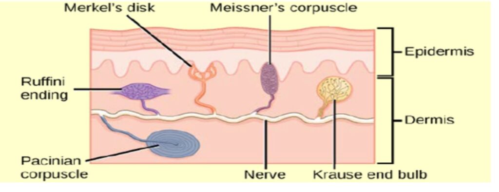
These are sensory receptors for the sensation of cold on the limbus, conjunctiva, skin and tongue. Sometimes referred to as “Krause’s end bulbs”

Krause ‘s membranes:

These are defined as isotropic bands in striated muscle fibre that consist of disks of sarcoplasm and connect the individual fibrils. Also known as Z-Disc or Dobie’s line.

Krause respiratory bundle:

A slender compact fibre bundle that is also known as the “solitary tract”. It is referred to as “Gierke respiratory bundle”, coined in honour of anatomist Hans Paul Bernhard Gierke.

Fun fact: Bacteriologist Robert Koch was Wilhelm Krause’s student at Gottingen

Read about “Alexander- The doctor who was shot by his patient

Alexander- The doctor who was shot by his patient

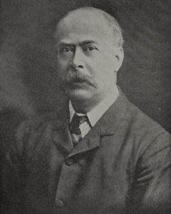
Professor Gustav Alexander was an Austrian Otolauringologist. Born in  Vienna in 1873. Prof. Alexander received his medical degree in 1898. He was mainly trained in anatomy, neurology, histopathology and other areas in addition to ontology. Today is his death anniversary.

He has been famous for his description of Alexander’s law.  His edict that gaze in the direction of the fast component increased the intensity of the nystagmus, while gaze in the opposite direction had the reverse effect, added diagnostic significance to the laws of nystagmus.

He personally funded and established a histopathology lab for his students. In addition to being a wonderful scientist, he was also a talented pianist. The expression of his appreciation for the arts extended to his collection of paintings, bronzes, silver and early tiles.

Later, he published Ear diseases of childhood and the handbook of Oto-neurology. On April 12th, 1932, Professor Gustav Alexander was shot dead by his own patient. He had performed saddle-nose surgery on a patient, Johann Sokoup, who was not happy with the results, hence, the same person has tried shooting 22 years before that and missed the aim, unfortunately. His death is considered an irreparable loss to the otology. He published an average of 12 articles every year.

References:

https://journals.sagepub.com/doi/pdf/10.1177/014556139607500307

https://www.cambridge.org/core/journals/journal-of-laryngology-and-otology/article/professor-gustav-alexander-vienna/632C779BD7824B1201CABD818452427F

Hassall and Hassall-Henle bodies

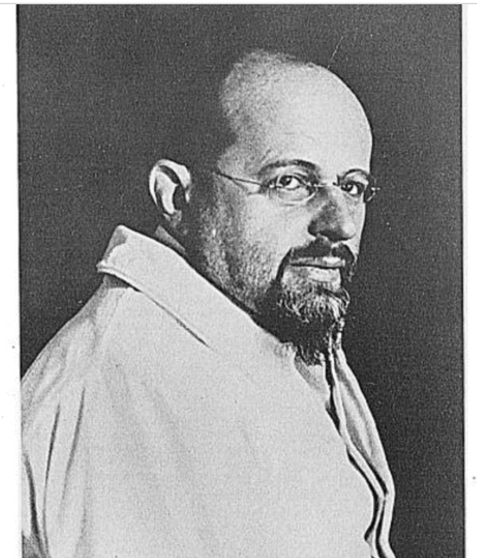
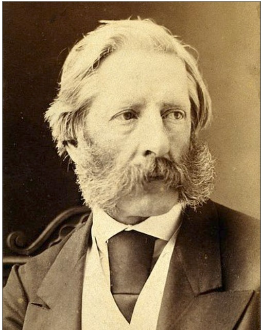
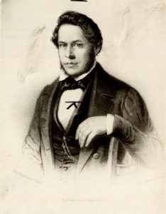
It is the death anniversary of Arthur Hassall Henle. (13th Dec 1817- 9th April,1894) He is a British physician, chemist and microscopist who is primarily known for his work in public health and food safety.

He published a two-volume study, The Microscopic Anatomy of the Human Body in Health and Disease, the first English textbook on the subject.

Are you curious to read about another scientist? Click on this link for more information

He became famous with his book, A microscopical examination of the water supplied to the inhabitants of London, which became an influential work in promoting the cause of water reform.

Two medical terms are named after Hassall: 

Hassall corpuscles- The spherical bodies in the medulla of thymus glands

Hassall-Henle bodies-small abnormal transparent growths on the posterior surface of Descemet’s membrane

The Thames caused the spread of many diseases, including cholera. In the early 1850s, he also studied food adulteration. His reports led to the first Food Adulteration Act of 1860. He continued his interest in climate and disease and published extensively on climatic treatments for TB.

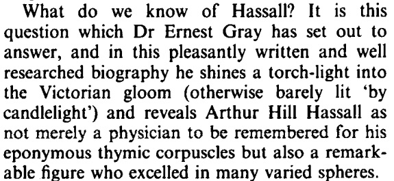
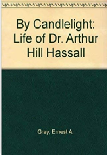
The biography of Hassall is written by Dr Ernest Gray.

What do we know of Hassall? It is this question which Dr Ernest Gray has set out to answer, and in this pleasantly written and well researched biography he shines a torch-light into the Victorian gloom (otherwise barely lit ‘by candlelight’) and reveals Arthur Hill Hassall as not merely a physician to be remembered for his eponymous thymic corpuscles but also a remarkable figure who excelled in many varied spheres

hassall

He died in 1894 at the age of 77.

References:

Nobel laureate- Allvar Gullstrand

Hello guys,

Here I am with another interesting article in the category Today in History on the one and only Ophthalmologist who has been awarded Nobel prize for Ophthalmology related research. Hope you have guessed it by now.

Allvar Gullstrand
This photograph of Allvar Gullstrand with his corneal microscope is reproduced from the British Journal of Ophthalmology 2010; 94:826

Yes, It’s Allvar Gullstrand and it’s his birthday. A Swedish Ophthalmologist and a self-taught mathematician. Slit-lamp invented by him is the most common inventions of him which every student must have known by now. When I sat down to write this article, I have found tons of information online. So, I will just try to make things as simple as possible.

Read More

Founder of Scientific Ophthalmology - Albrecht Von Graefe

Friedrich Wilhelm Ernst Albrecht Von Graefe (22 May 1828 – 20 July 1870)

Friedrich Wilhelm Ernst Albrecht Von Graefe

Happy Birthday to the founder of Scientific Ophthalmology and father of glaucoma- Von Graefe

He was born in Berlin on 22th May 1828 and was the third son of Carl Ferdinand Von Graefe. His father died when Graefe was only 12. Graefe was an extremely brilliant student from childhood. In 1847, he graduated at the age of 19.

Read More

Robert Walter Doyne

Today is the birthday of Robert Walter Doyne.

Born on May 15, 1857, and was the 2nd son of late Reverend P. W. Doyne. Educated at Bristol medical school and St. George hospital in London. He then entered the medical service of the Navy. But after marriage in 1885, he had settled in Oxford, devoting himself to Ophthalmology.

Robert Walter Doyne
Robert Walter Doyne

At that time, there was neither an Ophthalmic surgeon nor a clinic in the city. After many difficulties, he founded Oxford Eye Hospital. He was an extraordinary clinical observer and some of his observations are given below.

Doyne’s cataract (discoid cataract):

In 1906, Nettleship and Ogilvie published an extensive study of the occurrence of congenital cataract in the Coppock family. The first observation on this familial anomaly was made by Doyne, who had the opportunity of examining the first, second and third, and possibly fourth, cases in this family between 1888 and 1896. Members of the family were affected by a definite and peculiar type of stationary congenital cataract, which showed but slight variation in clinical appearance. It was a disk-shaped opacity of steel grey colour in focal light. Furthermore, opacity made the fundus invisible or, at best, only dimly discernible.

Doyne choroiditis (genetic trait resulting in retinal degeneration and retinal drusen):

In 1899, he first described an English family with a condition which he called choroiditis, and which was subsequently known as Doyne’s honeycomb degeneration of the retina. His first description was brief and stressed the familial nature of the condition. Choroiditis noted in four sisters and their father and one of his brothers and the paternal grandmother. There was a honeycomb appearance of the fundus with white spots affecting the macular region of the disc. Eleven years later, a female cousin of the same family was also affected. He thought of making copies of the pedigree and circulating to his colleagues as the family was dispersed all over the country. There were two families which he was trying to link together: Buckinghamshire and Oxfordshire.

Also read Brain behind bunch of eponyms.

Doyne’s conjunctivitis (a form of conjunctive blepharitis), Doyne’s honeycomb macular dystrophy, and Doyne Iritis (grey precipitate found on iris), and also invented a number of appliances including, stereoscopes, tonometers and retinoscopes. He was the first person to describe small breaks in Bruch’s membrane. Also called Angiloid streaks (Knapp streaks or Knapp striae) by Herman Jakob Knapp after few years. He also described pseudo-cataract (so-called lens with double focus). Wrote “Notes on the more common diseases of the eye”.

He2fhref="https://livesonline.rcseng.ac.uk/client/en_GB/lives/search/detailnonmodal/ent:$002f$002fSD_ASSET$002f0$002fSD_ASSET:373633/one?qu=%22rcs%3A+E001450%22&rt=false%7C%7C%7CIDENTIFIER%7C%7C%7CResource+Identifier">2fSD_ASSEThref="https://livesonline.rcseng.ac.uk/client/en_GB/lives/search/detailnonmodal/ent:$002f$002fSD_ASSET$002f0$002fSD_ASSET:373633/one?qu=%22rcs%3A+E001450%22&rt=false%7C%7C%7CIDENTIFIER%7C%7C%7CResource+Identifier">2f0href="https://livesonline.rcseng.ac.uk/client/en_GB/lives/search/detailnonmodal/ent:$002f$002fSD_ASSET$002f0$002fSD_ASSET:373633/one?qu=%22rcs%3A+E001450%22&rt=false%7C%7C%7CIDENTIFIER%7C%7C%7CResource+Identifier">2fSD_ASSET:373633/one?qu=%22rcs%3A+E001450%22&rt=false%7C%7C%7CIDENTIFIER%7C%7C%7CResource+Identifier"> died at his residence in Woodstock Road, Oxford, on August 30th, 1916, leaving a widow and two sons, of whom one - Philip Geoffrey Doyne, FRCS. Two years after his death, Doyne memorial lecture and medal was established which even today remains as one of the top awards in British Ophthalmology.

Brain behind bunch of eponyms - Henle

Crypts of Henle (Microscopic pockets located in the conjunctiva of the eye), Henle’s membrane (Bruch’s layer forming the inner boundary of the choroid of the eye), Hassall-Henle bodies, Henle’s fissure, Henle’s ampulla, Henle’s layer, ligament, Spine, Henle’s sheath, loop of Henle, Glands and tubes of Henle

Do you realize what’s common in all the above?

Friedrich Gustav Jakob Henle

Jacob Henle was a great German anatomist and one of the most important histologists of all times. He was born on July 19, 1809, Fürth, near Nuremberg, Bavaria Read More

Mastermind behind Diopter

Do you know who coined the term Diopter?

Most of us would have guessed it to be a physicist. But to our surprise, he was an Ophthalmologist. Yes, he was the pioneering eye doctor, Dr.Ferdinand Monoyer and it’s his birthday today.

Ferdinard Monoyer - Diopter
Ferdinard Monoyer

Read More